
Figure 3. AMCase induces Akt phosphorylation.
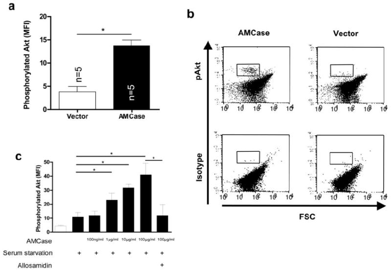
a. Intracellular phosphorylated Akt was analyzed by flow cytometry (upper panel) or Western blotting (lower panel) in A549 cells transfected with the empty vector (Vector) or with human AMCase (AMCase). Phosphorylated Akt (p-Akt) is shown in relation to total Akt (Akt-total). * p<0.05, Mann-Whitney U test
b. Representative FACS dot-plots of intracellular levels of phosphorylated Akt or the corresponding isotype control are shown in A549 cells transfected with the empty vector (Vector) or with human AMCase (AMCase). The quadrant gate indicates the positive Akt staining.
c. Intracellular phosphorylated Akt was analyzed by flow cytometry in A549 cells transfected with the empty vector (Vector) or with full-length human AMCase (AMCase). Data from three independent experiments is shown. * p<0.05, Mann-Whitney U test