
Figure 1.
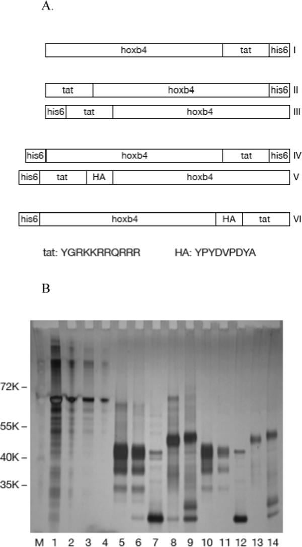
Expression of recombinant human hoxb4. Diagram of six versions of recombinant hoxb4 with different locations of the histidine and tat tags (A). E. coli expressed, ni-affinity chromatography and HiTrap SP HP ion-exchange chromatography purified hoxb4 proteins were analyzed by SDS-PAGE and stained with Silver Quest staining kit. Lanes 1, 2, 3 and 4: BSA 1 μg, 0.2 μg, 0.1 μg and 0.02 μg; lanes 5, 6, 7, 8 and 9: version I, II, IV, V and VI hoxb4 each 0.6 μg (3ul) before endotoxin removal; lanes 10, 11, 12, 13 and 14: version I, II, IV, V and VI hoxb4 each 3ul after endotoxin removal (B).