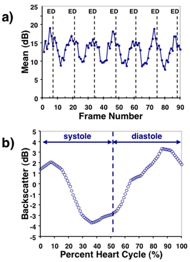
Figure 2.
a) Example of the measured cyclic variation of backscatter expressed in dB for 6 heart cycles from the left-ventricular free wall of one of the fetuses with the end-diastolic frames indicated by “ED”. b) Corresponding composite cyclic variation curve. The average systolic and diastolic intervals are indicated.

