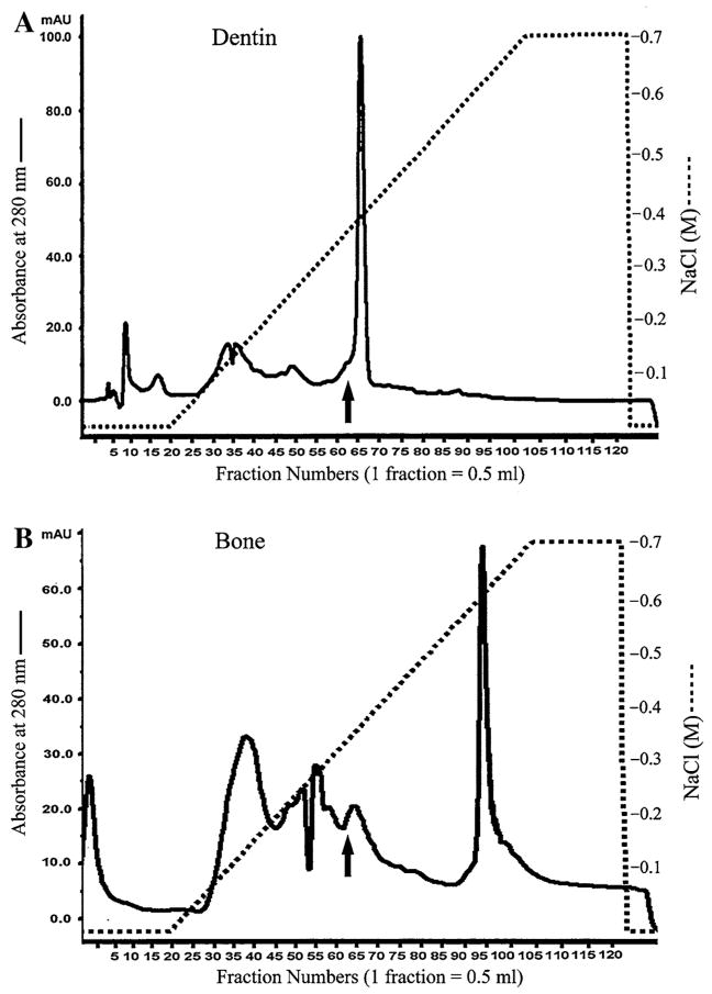
Fig. 1.
Separation of NCPs from the ECM of rat dentin and bone. (A) The Q-Sepharose ion-exchange chromatogram of ES1 of the dentin extract. Arrow indicates the fraction that contained DMP1 and its fragments and was used for Stains-All staining and Western immunoblotting in Fig. 2. (B) The Q-Sepharose ion-exchange chromatogram of ES1 of the bone extract. Arrow indicates the fraction used for Stains-All staining and Western immunoblotting in Fig. 2

