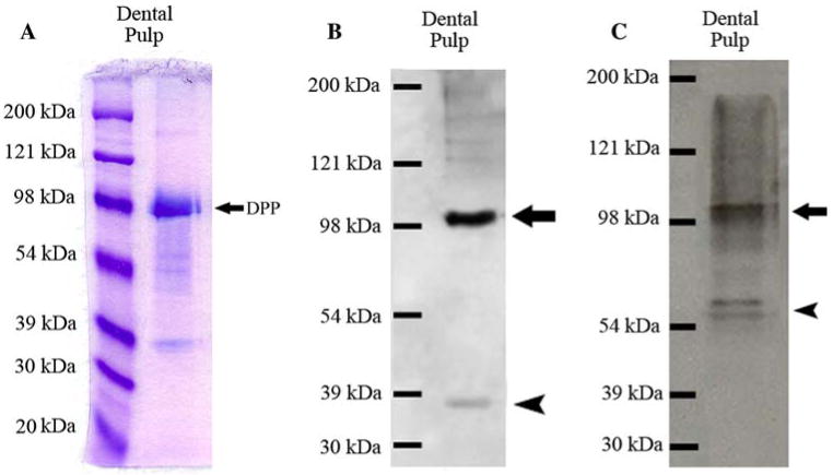
Fig. 3.
Detection of the full-length form of DMP1 and its processed fragments in the extract of rat dental pulp. (A) Stains-All staining for a Q-Sepharose chromatographic fraction of dental pulp extract. The sample was from a chromatographic fraction of the Gdm-HCl extract of the dental pulp (corresponding to fraction 63 of the Q-Sepharose chromatograms of dentin or bone extracts). Sixty microliters of sample in 6 M urea solution was loaded. The strong blue band migrating just below 98 kDa represents DPP (arrow). DMP1 fragments are visible below the DPP band. The protein band representing the full-length form of DMP1 cannot be observed between 98 and 121 kDa, due to the relatively small amounts of sample loaded. (B) Western immunoblotting using monoclonal anti-DMP1-N-9B6.3 antibody. Note that both the full-length form of DMP1 (arrow) and its processed NH2-terminal fragment (arrowhead) are present in the extract of the pulp/odontoblast complex. (C) Western immunoblotting using polyclonal anti-DMP1-C-785 antibody. Both the full-length form of DMP1 (arrow) and its processed COOH-terminal fragment (arrowhead) are present in the extract of pulp/odontoblast complex

