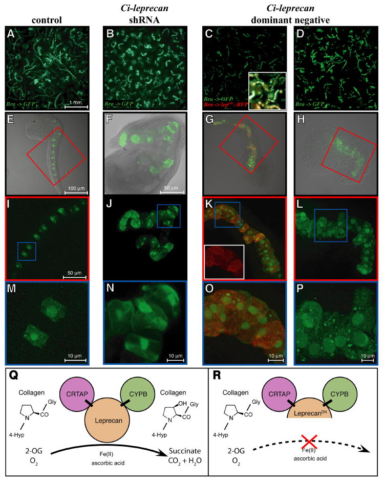
Figure 6. Analysis of the notochord phenotypes induced by the over-expression, dominant-negative and knock-down of Ci-leprecan.
Comparative microscopic study of Ciona embryos electroporated with the Ci-Bra->GFP reporter alone (A,E,I,M) or in combination with either of the following constructs: shLEP-767 (B,F,J,N), Ci-Bra->Ci-leprecanDN∷RFP (C,G,K,O) and Ci-Bra->Ci-leprecanDN (D,H,L,P). (A-D) Embryos photographed at low magnification using an upright microscope. (E-P) Collapsed image stacks obtained with a confocal microscope. The inset in (C) shows a small area of the same image with the red and green channels merged. (E-H) Merge of the bright-field and dark-field images of representative individual embryos. (I,K,L) Dark-field high-magnification images of the areas boxed in red in panels E,G,H and (J) dark-field high-magnification view of the whole embryo shown in (F). The inset in (K) shows the red channel image of the area boxed in blue. (M-P) Dark-field high-magnification images of the areas boxed in blue in panels I-L. (Q,R) A simplified model for the mechanism of action of the native Ci-Leprecan protein (Q) and of the dominant-negative protein employed in this study (R). The dominant-negative Ci-Leprecan lacks the iron-binding region of the P4Hα domain, hence it is not expected to be able to modify collagen, but rather to interfere with the native Ci-Leprecan by sequestering its collagen substrate and its binding partners, CRTAP and CYPB. Abbreviations: 4-Hyp = 4-hydroxyproline; 2-OG = 2-oxoglutarate.

