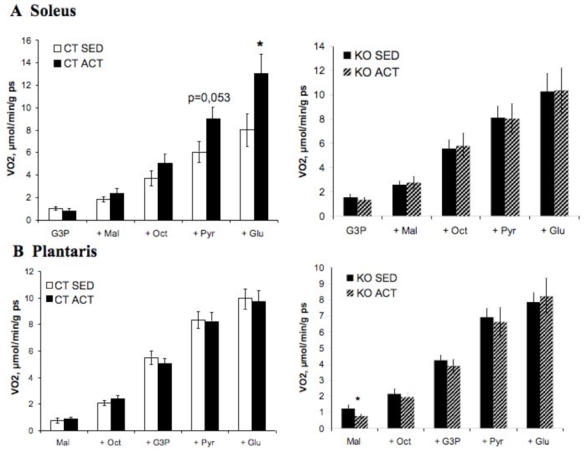
Figure 4. Role of AMPKα2 in skeletal muscle mitochondrial function.

Substrate utilization by mitochondria were determined in permeabilized fibers of soleus (A) and plantaris (B) muscles of sedentary (SED) and active (ACT) AMPK 2 −/− (KO) mice and littermate (CT) controls. Respiration rates were measured during the cumulative addition of substrates in saponin-skinned cardiac fibers of control and AMPKα2−/− mice. VO2: rate of O2 consumption in μmol·min−1·g dw−1. G3P: glycerol-3-phosphate, Mal: malate, Oct: octanoylcarnitine, Pyr: pyruvate, Glu: glutamate. * p<0.05 vs. control mice.
