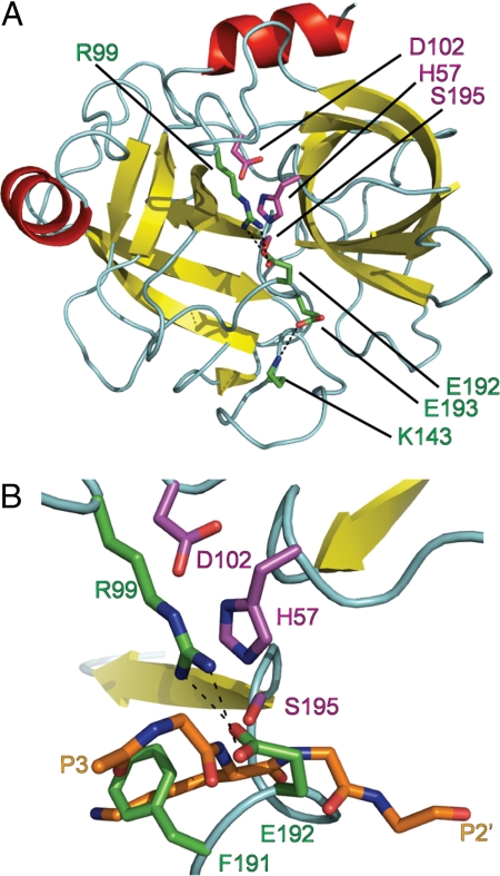
Fig. 1.
Granzyme C active site is occluded. (A) Diagram of granzyme C showing the chymotrypsin-like fold. (B) Enlarged view of the active-site cleft. Granzyme C was superposed with trypsin in complex with BPTI PDB ID code 2PTC). BPTI residues 13–17 indicate likely positioning of substrate P3–P2′. Cyan, loops and turns; red, α-helices; yellow, β-strands. Catalytic residues (magenta) D102, H57, and S195, inactivating residues (green) R99, K143, P191, E192, and E193 and BPTI main chain and P1 side chain residues (orange) are shown as stick models colored by element: red, oxygen; blue, nitrogen. The figures were rendered with PyMOL (32).

