Fig. 2.
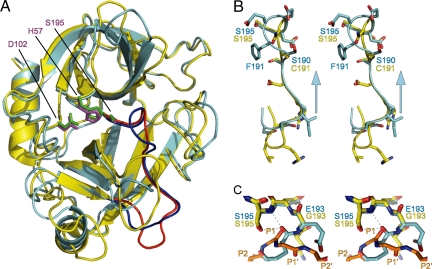
Granzyme C inactivation is caused by a register shift. (A) Superposition of granzyme C (cyan) with human granzyme A (yellow; PDB ID code 1ORF). The active-site triads are shown as stick models: magenta, granzyme C; green, granzyme A. The blue (granzyme C) and red (granzyme A) regions are enlarged in B. (B) Stereoview of the register shift in granzyme C. The arrow indicates the direction of register shift. (C) Stereoview of the oxyanion hole. Trypsin in complex with BPTI (PDB ID code 2PTC) was added to the superposition. BPTI residues 13–17 indicate likely positioning of substrate P3–P2′. Dashed lines indicate hydrogen bonds between granzyme C E193 carbonyl and S195 amide and Oγ. The figures were rendered with PyMOL (32).

