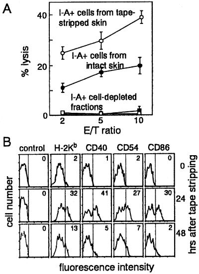
Figure 6.
Augmentation of CTL priming capacity of epidermal LCs by barrier disruption. (A) Ia+ cell-enriched and -depleted epidermal cells were prepared from tape-stripped (○) or intact (●) earlobes by separation with anti-Iab mA-conjugated magnetic beads. Cervical lymph node cells (1 × 10⁵) from intact mice were primed in vitro with TRP-2-pulsed epidermal cell populations (1 × 10⁴) at a ratio of 20 lymph node cells to 1 epidermal cell. Cytotoxicity was assayed by using TRP-2-pulsed LKb target cells at indicated effector-to-target (E/T) ratios. Data are expressed as mean ± SE of the results of two independent experiments. (B) Epidermal cells prepared from intact earlobes (0 hr) or those tape-stripped 12, 24, or 48 hr before were incubated with FITC-conjugated H-2Kb (AF6–88.5), CD40 (HM40–3), CD54 (3E2), CD86 (GL-1), or control mouse IgG mAb and phycoerythrin-conjugated anti-Iab mA (AF6–120.3). By flow cytometry, strongly Iab+ cells, representing LCs, were gated, and H-2Kb, CD40, CD54, or CD86 expression was visualized as a histogram. Numbers indicate percentage of LC populations that express H-2Kb, CD40, CD54, and CD86.

