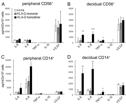
Fig. 2.
HLA-G homodimer induced cytokine protein secretion by human peripheral and decidual CD14+ macrophages and CD56+ NK cells. Peripheral CD56+ (A), decidual CD56+ cells (B), peripheral CD14+ cells (C), or decidual CD14+ cells (D) were cocultured in a 1:1 ratio with HLA class I-negative 721.221 cells (white bars), 721.221 cells transfected to express HLA-G monomer (gray bars), or 721.221cells transfected to express HLA-G homodimer (black bars). Supernatants were collected after 24 h for CD14+ macrophages or after 48 h for CD56+ NK cells. The concentration of cytokines (IL-6, IL-8, IL-10, TNFα, or VEGF) in each supernatant was measured by a multiplex cytokine assay. The SD was calculated from the mean of 4 experiments, each with an individual donor. Statistical significance: *, P < 0.05; **, P < 0.01; and ***, P < 0.001. Note that the scale of C is compressed ≈4-fold relative to D because the secretion of IL-8 by peripheral CD14+ cells is so much larger than that of decidual CD14+ cells.

