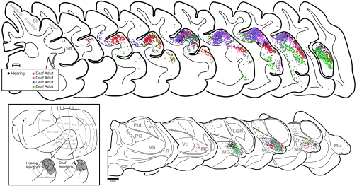
Fig. 4.
Anatomical tracer injected into A1 revealed the same pattern of connections in both adult-deafened (≈76 d duration) and hearing ferrets. (Inset) Lateral view of ferret cortex with expanded views of the auditory cortices depicting the tracer deposits (gray) for hearing and deafened animals. (Top) Serially arranged coronal sections containing somatosensory and auditory cortices. Each dot represents 1 labeled neuron (black dots represent 1 hearing adult ferret; colored dots represent 4 deafened adult ferrets). The distribution of black and colored dots is essentially co-extensive; areas of somatosensory cortex (S1; SII) are largely devoid of label. No labeled neurons were identified in sections anterior or posterior to those depicted. (Bottom) Serially arranged sections through thalamus with the auditory (MG, medial geniculate), somatosensory (Vb, ventrobasal), visual (LGN, lateral geniculate; Pul, pulvinar), and non-specific (PO, posterior; LP, lateral posterior) nuclei depicted. Each dot represents 1 labeled neuron (black dots, hearing; colored dots, deafened). The distribution of black and colored dots is essentially co-extensive within the MG; somatosensory thalamus (Vb) is devoid of label in both conditions.

