
Figure 3.
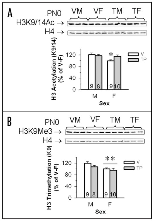
Testosterone propionate (TP) increased H3K9/14Ac, but not H3K9Me3 in the cortex/hippocampus of female mouse pups. (A) Top shows a set of representative immunoblot from PN0 males and females. The western blots on the top row show H3K9/14Ac bands and the bottom row is total H4 from the same individuals. Mean optical densities (±SEM) of H3K9/14Ac in the cortex/hippocampus of vehicle (V) and TP-treated male (M) and female (F) neonates were normalized to levels of H4, and expressed as the percentage of V-treated F controls (100%). *Females whose dams received vehicle during gestation had significantly lower levels of H3K9/14Ac as compared with the other three groups (p < 0.05). (B) Top shows a representative immunoblot from PN0 males and females. The western blots on the top row show H3K9Me3 bands and the bottom row is total H4 from the same individuals. Mean optical densities (±SEM) of 3mH3 in the cortex/hippocampus of V and TP-treated M and F neonates were normalized to levels of H4, and expressed as the percentage of V-treated females (as 100%). **Females have significantly less H3K9Me3 than males, p < 0.05. Sample sizes are shown in each histogram.