
Figure 3. IL-6 produced by bone marrow derived cells is required for CAC tumorigenesis.
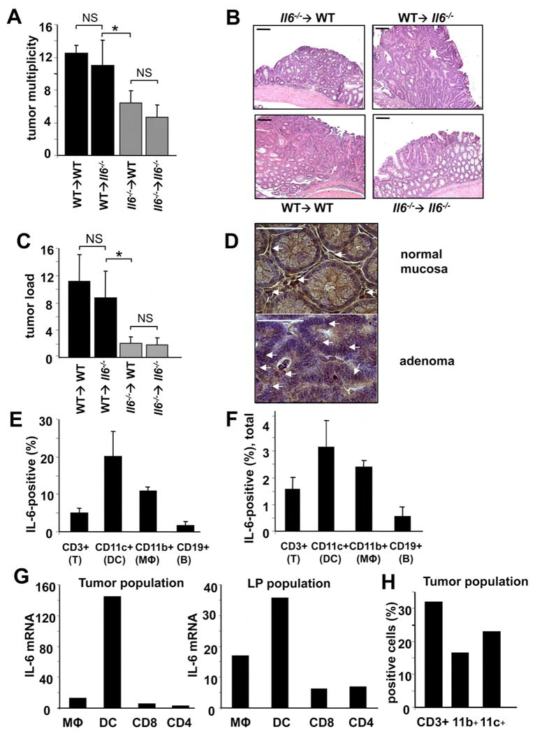
(A) Tumor multiplicity in radiation chimeras subjected to induction of CAC using 2.5% DSS. Results are averages ± s.d. (n>8), * p<0.01. (B) Paraffine embedded sections of adenoma-containing colons stained with H&E. Scale bar- 100 μm. (C) Average tumor loads in radiation chimeras. Results are averages ± s.d. (n=5), NS- not significant, * p<0.01. (D) Immunohistochemical analysis of IL-6 expression in DSS-treated colons (normal mucosa) or CAC-bearing colons of WT mice. Scale bar- 50 μm. (E,F) Intracellular IL-6 cytokine staining of PMA+ionomycin restimulated tumor infiltrating cells analyzed by flow cytometry. Results are averages ± s.d. (n=3). (E) Percentages of IL-6 expressing cells in each given population (DC, macrophages, T and B cells) (F) Percentages of each population positive for IL-6 cells among all tumor infiltrating cells. (G) Macrophages (CD45+CD11b+CD11c+), dendritic cells (CD45+CD11b+CD11c+) and T cells (CD45+CD4+ or CD45+CD8α+) isolated by FACS sorting were analyzed for IL-6 mRNA by Q-RT-PCR. Tumor population- tumor infiltrating cells from pooled tumors. LP population- lamina propria cells from colons from which the tumors were excised. (H) Percentage of macrophages (CD11b+), T cells (CD3+) and dendritic cells (CD11c+) in total lamina propria cells isolated from pooled CAC tumors from WT mice.