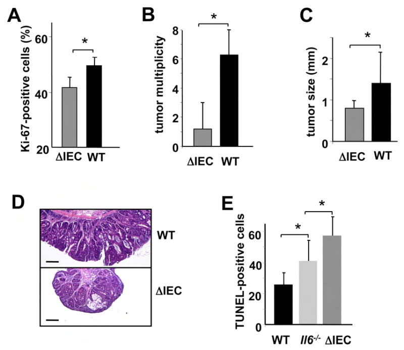
Figure 6. STAT3 is critical for CAC tumorigenesis.

(A) Percentage of Ki-67-positive cells in WT and Stat3ΔIEC colonic crypts 10 days after initiation of DSS exposure. Results are averages ± s.d. (n=3). * p=0.03. (B) Tumor multiplicity in WT and Stat3ΔIEC mice subjected to induction of CAC. Results are averages ± s.d. (n=8), *p=0.004. (C) Tumor sizes in WT and Stat3ΔIEC mice. Results are averages ± s.d. (n=8), * p=0.012. (D) Paraffine embedded sections of adenoma-containing colons of WT and Stat3ΔIEC mice were stained with H&E. Scale bar- 100 μm. (E) Apoptosis in colons of DSS treated mice was evaluated on day 4 after DSS administration by TUNEL staining. Amount of TUNEL-positive cells per microscope field was determined. Results are averages ± s.d. (n=3). * p=0.05.
