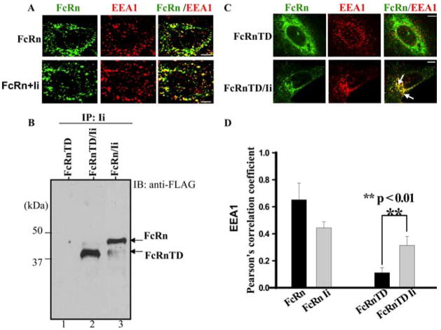
FIGURE 5.
The Ii chain redirects tailless FcRn-TD to the early endosome. Bars, 5 μm. A, Endocytic trafficking of FcRn in HeLaFcRn and HeLaFcRn + Ii transfectants. HeLaFcRn cells were transiently transfected with pBUDCE4 or pBUDCE4-Ii vector. Cells were fixed, permeabilized, and costained for FcRn (green) or early endosomal marker EEA1 (red). Puncta that appear yellow in the merged images (right) indicate colocalization of FcRn with the EEA1. B, Association of tailless FcRnTD with the Ii chain. The cell lysates from HeLaFcRnTD (lane 1), HeLaFcRnTD/Ii (lane 2), and HeLaFcRn/Ii (lane 3) were immunoprecipitated by anti-HA mAb. The immunoprecipitates were subjected to Western blot. Immunoblots (IB) were blotted with rabbit anti-FLAG Ab and HRP-conjugated goat anti-rabbit Ab. The blot was developed with ECL. C, Immunofluorescence analyses of HeLa cells expressing either tailless FcRn-TD alone (top panel) or FcRn-TD and Ii (bottom panel). Transfected cells were fixed, permeabilized, and stained with an Ab to FLAG (green) or EEA1 (red). Arrows, Colocalization (yellow) of the proteins. D, Averages of the colocalization coefficients in A and C. Pearson’s correlation coefficient were calculated. For each experiment, 15 cells were analyzed in 3 different optical regions.

