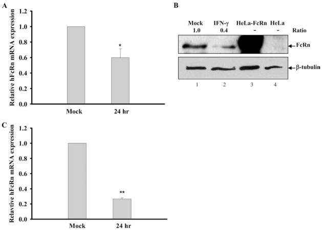
FIGURE 2.
Down-regulation of human FcRn expression in THP-1 cells and human PBMCs by IFN-γ. A and C, Effect of IFN-γ treatment on FcRn mRNA expression in THP-1 and PBMCs. The macrophage-like THP-1 cells (A) or freshly isolated human PBMCs (C) were treated with or without IFN-γ (25 ng/ml) for 24 h. The levels of FcRn mRNA were measured by quantitative real-time RT-PCR analysis as described in Materials and Methods. Data are mean ± SD of three independent experiments. *, p < 0.05; **, p < 0.01. B, Western blot analysis of FcRn expression in THP-1. The cell lysates (20 μg) from THP-1 (lane 1), IFN-stimulated THP-1 (lane 2), HeLa-FcRn (lane 3), and HeLa (lane 4) were subjected to 12% SDS-polyacrylamide gel electrophoresis. The proteins were transferred to nitrocellulose membrane and blotted with FcRn- (top panel) or β-tubulin-specific Ab (bottom panel). Blots were then incubated with anti-IgG-HRP and visualized with the ECL method. The ratio of the mock was assigned a value of 1.0, and the values from other groups were normalized to this value. The ratios of FcRn- and β-tubulin are shown above the lanes.

