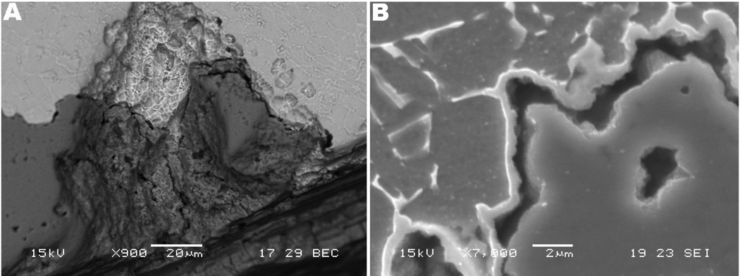
Fig. 8.
SEM micrograph of cracking in the Z component. (a) BS image showing presence of microstructure (top) and debris layer (darker gray, bottom) (900×). (b) Note that the gray (darker gray, bottom) debris layer fills the region of a pit and appears to be a reaction layer converting the alloy to debris with very close proximity with the alloy topography (7,000×).

