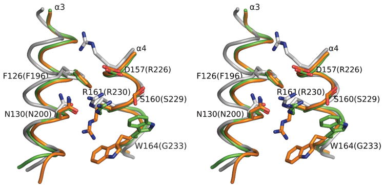
Figure 7.
Arginine fingers of Hda. The superimposed conformations of the two arginine fingers (Arg161) in the dimer and surrounding conserved residues of Hda (orange and green). The positional equivalent residues of DnaA (PDB id 2hcb, gray) are also shown. The corresponding residue numbers for DnaA are shown in parenthesis.

