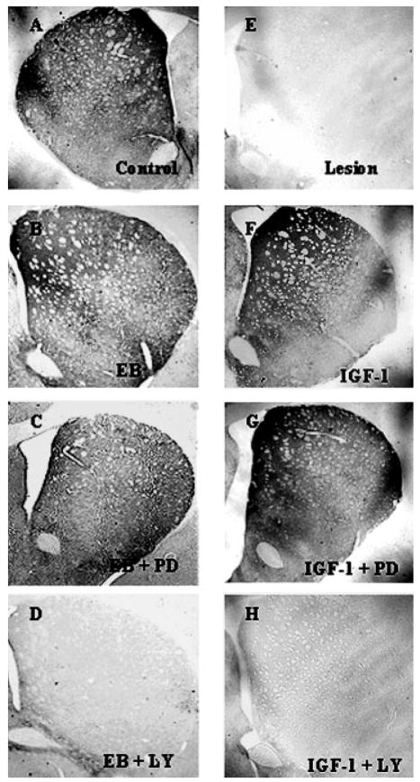
Figure 4.
Photomicrographs of striatal TH immunoreactivity 7 days after unilateral injection of 6-OHDA (2.0 μL of 4 μg/μL) into the medial forebrain bundle. A: Nonlesion control, (E) Lesion side. B: 17β-estradiol benzoate (EB; 20 μg) was given 24 h prior to lesion, (C) EB + PD98059 (MAPK/ERK inhibitor; 0.10 μg/μL) and (D) EB + LY294002 (PI3K/Akt inhibitor; 0.10 μg/μL). For IGF-1 treated groups (F–H). F: Central infusion of IGF-1 (100 μg/mL), (G) IGF-1 + PD98059 (0.10 μg/μL) and H) IGF-1 + LY294002 (0.10 μg/μL). IGF-1 and inhibitors were continuously infused for 7 days.

