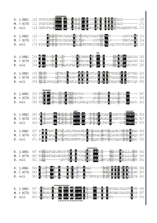
Figure 4.
Partial view of amino acid sequence alignment of glucan phosphorylases. The identical amino acids in the three sequences are indicated by black background. The deduced active sites are indicated by lines, and the putative pyridoxal-phosphate cofactor binding site is indicated by asterisks.

