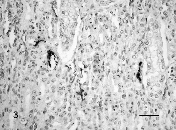
Fig. 3.
Kidney; Leptospira-infected equine fetus No. 3. Typical leptospiral wavy forms and aggregates in the lumen of several tubuli. Leptospires are detected with LipL32-specific antiserum and horseradish peroxidase–labeled polymer (EnVision+Kit). The section is counterstained with Mayer’s hematoxylin. Bar = 60 μm.

