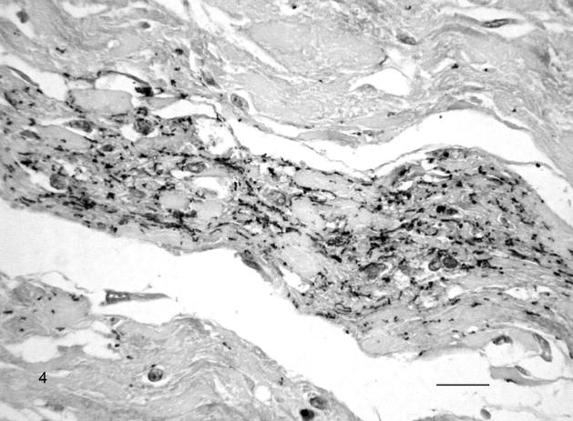
Fig. 4.
Chorioallantoic membrane; Leptospira-infected equine fetus No. 1. Large amount of extracellular leptospiral wavy forms, small cocci, and aggregates in the allantoic connective tissue. Leptospires are detected with multivalent Leptospira antiserum, and horseradish peroxidase–labeled polymer (EnVision+ Kit). The section is counterstained with Mayer’s hematoxylin. Bar = 60 μm.

