
Figure 2. Treatment with SERMs inhibits proliferation of ER-α deficient splenocytes.
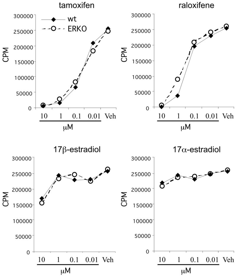
Splenocytes from naïve female estrogen receptor-alpha knockout (ERKO-alpha) mice were stimulated with plate-bound anti-CD3/anti-CD28 and treated with the indicated amount of tamoxifen, raloxifene, 17β-estradiol, or 17α-estradiol for 72hrs. [3H]-thymidine was added for the last 18 hrs of culture and the incorporation of radioactive thymidine by the cells was quantified as a measure of cellular proliferation. The data are presented as mean counts per minute from triplicate cultures.