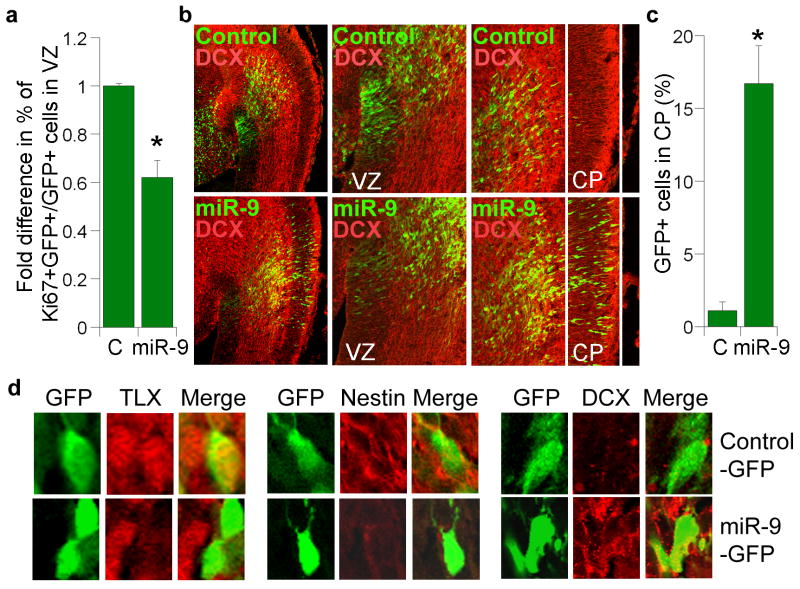
Fig. 5. In utero electroporation of miR-9 in embryonic neural stem cells.
a. In utero electroporation of miR-9 decreased cell proliferation in the ventricular zone (VZ) of embryonic brains. Proliferating cells were labeled by Ki67. Percent of Ki67-positive cells out of GFP-positive cells (Ki67+GFP+/GFP+) in miR-9-electroporated brains was calculated and normalized with the percent of Ki67-positive cells in control RNA (C)-electroporated brains. s.d. is indicated by error bars. * p=0.002 by Student's t-test. b. Electroporation of miR-9 led to precocious outward cell migration. The transfected cells were shown green due to the expression of GFP marker. Control: control RNA; DCX: double cortin; VZ: ventricular zone; CP: cortical plate. The left panels are 10× images, the middle and right panels are 20× images. c. Quantification of control RNA (C) and miR-9-electroporated cells (GFP+ cells) that migrated to the cortical plate (CP). Error bars are s.d. of the mean. * p=0.02 by Student's t-test. d. Immunostaining of cells from control-GFP or miR-9-GFP-electroporated brains.

