
Figure 8.
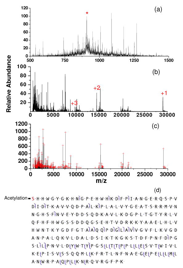
(a): Beam-type CID of carbonic anhydrase [M+32H]32 (KE=588.8 eV, Q2 LMCO = 600) (b): Simultaneous beam-type CID and ion/ion reaction (anion injection time = 100 ms) (c): Deconvoluted spectrum of (b). Peaks selected by the peak picking program are indicated by red crosses. Panel (d) shows the fragmentation pattern in spectrum (b) identified by ProSight PTM 2.0. (‘*’in (a) represents signal from the residual carbonic anhydrase [M+32]32+ precursor ion. The numbers in (b) represent charge states of the residual precursor ion that have resulted from ion/ion proton transfer reactions.)