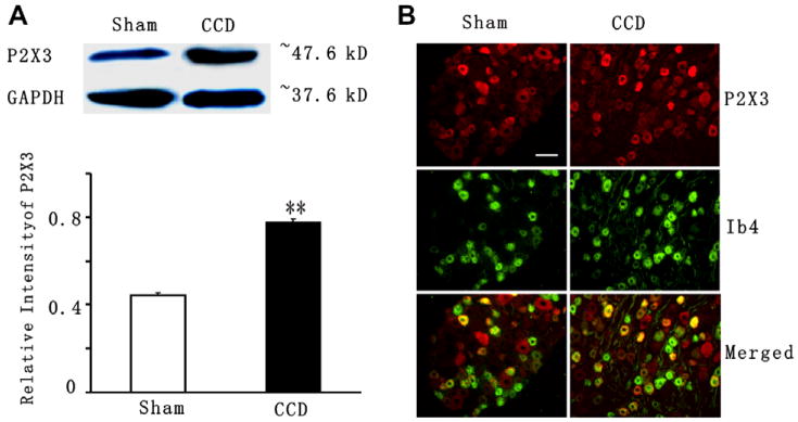
Fig. 3.
Up-regulation of P2X3 receptor in the CCD-injured DRG. (A) Western blots of DRG protein extracts probed for P2X3. GADPH serves as the loading control. P2X3 levels (normalized with density of GADPH bands) increase in the DRGs ipsilateral to the CCD surgery (16 DRGs in 4 CCD groups with 2 rats in each group) compared with DRGs ipsilateral to the sham operation (12 DRGs in 3 sham group with 2 rats in each group) (**p < 0.01, student’s test). (B) P2X3 receptor (red) and IB4 (green) immunoreactivity in L4/L5 DRG in the sham-operated rats (Left) and after CCD (right). Nerve fibers as well as cell bodies were stained for IB4 after CCD (right). Note that an increased number of neurons showed colocalization of P2X3 receptors and IB4 in the CCD model (yellow, right) as compared to in the sham (yellow, left). Scale bar = 100 μm.

