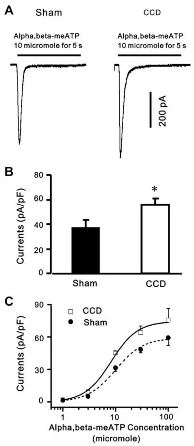
Fig. 5.
α,β-meATP-evoked currents are potentiated in DRG neurons after CCD. (A) whole-cell voltage-clamp recordings were made to record α,β-meATP-evoked currents from DRG neurons from sham-operated and CCD groups. (B) α,β-meATP (10 μM) evoked larger inward currents in CCD neurons than in sham-operated neurons at a holding potential of −60 mV (*p < 0.05, n = 17, unpaired t-test). (C) Dose–response curves for α,β-meATP-evoked fast responses. Dose–response curves were constructed as a function of α,β-meATP concentration by averaging α,β-meATP-evoked peak amplitudes from DRG neurons from control and CCD rats, respectively. The data points were obtained from 8 to 20 neurons. The α,β-meATP EC50 values were 9.95 μM in control neurons and 8.23 μM in CCD neurons. The changes in α,β-meATP affinities for P2X receptors in CCD neurons were not significant.

