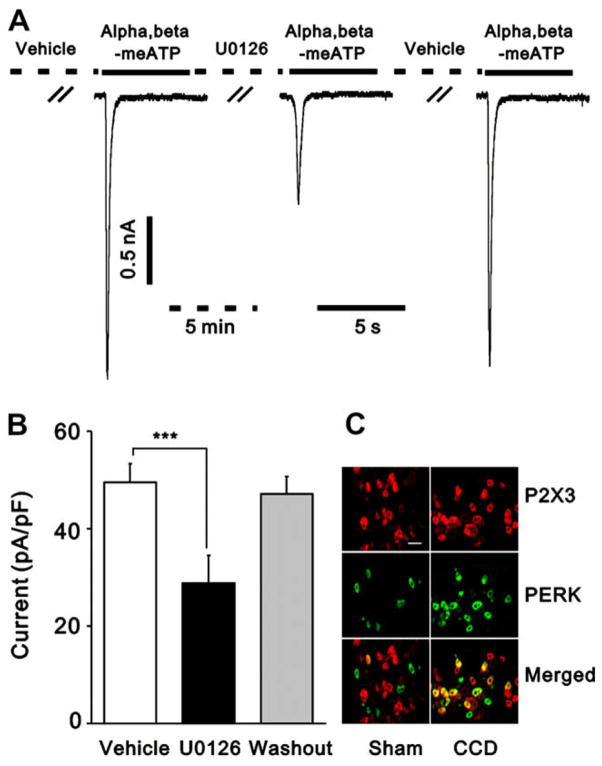
Fig. 6.
pERK mediated the enhancement of α,β-meATP-induced currents in CCD neurons. (A) representative current traces showing an extracellular application of a specific inhibitor for the mitogen-activated/ERK kinase pathway, U0126 (10 μM), for 5 min greatly reduced α,β-meATP-induced current in a CCD neuron. Currents were recorded at a holding potential of −60 mV. The effect of U0126 was reversible. (B) The mean amplitude of α,β-meATP-induced current was significantly lower after U0126 treatment than before U0126 application (***p < 0.001, paired t-test). (C) ERK was activated in P2X3-positive DRG neurons. More P2X3 -IR (red) or pERK-IR (green) neurons were seen in CCD-injured DRG (right) than in sham-DRG (left). The merged images (yellow) of P2X3 -IR and pERK-IR from the same section show colocalization of pERK-IR and P2X3-IR in DRG neurons in a CCD rat (right) and a sham-operated rat (left). A few double-labeled neurons (yellow) were seen in DRG neurons of the CCD rat but rarely found in the sham-operated rat. Scale bar = 100 μm.

