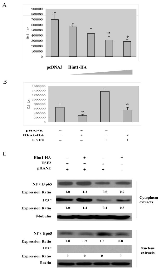
Figure 5. HINT1 inhibits NF-κB transcription factor activity in HepG2 cells.
(A) and (B), pNF-κB-Luc (400 ng/well) and the CMV-β-gal reporter control plasmid DNAs, together with pHA-Hint1 (0, 0.5, 1.0, 1.5, 2.0 μg) in (B) or pHA-Hint1 (1.0 μg) with our without CMV-USF2 (1.0 μg) plasmid DNAs, as indicated, were transfected into the HepG2 cells. Thirty-six hours later luciferase activity was assayed as described in Fig. 3A. (C), pHA-Hint1 or the CMV-USF2 plasmid DNAs were transfected into HepG2 cells, as indicated. At 48 hours post transfection, cytoplasmic and nuclear proteins were extracted, and each fraction was analyzed by Western blots to detect the expression levels of p65, IκBα, β-tubulin and β-actin. The latter two proteins were used as loading controls for the cytoplasmic and nuclear extracts, respectively. Expression ratios were calculated after normalization for β-tubulin and β-actin. Assays were repeated three times and gave similar results.

