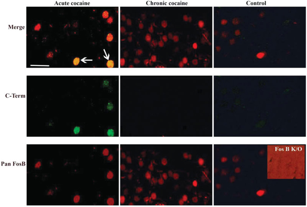
Fig. 1.
Double-labeling fluorescence immunohistochemistry using the anti-FosB (pan-FosB, Santa-Cruz) or anti-FosB (C-terminus) antibody through the nucleus accumbens of animals treated with acute or chronic cocaine and a control rat. The pan-FosB antibody stains both ΔFosB and FosB-positive nuclei (note nuclei from acute cocaine treatment stain with this antibody in green, and double stain with both antibodies “merge” row), whereas the C-terminus antibody stains FosB-positive nuclei only (note the absence of labeling in chronic cocaine tissue labeled with this antibody). All nuclei in the chronic cocaine-treated tissue are ΔFosB positive as they are stained only with the pan-FosB antibody and not with the C-terminal antibody. The inset shows a nucleus accumbens section from a fosB knockout mouse stained with the pan-FosB antibody; the lack of staining demonstrates the specificity of the antibody for fosB gene products. The background on this panel is intensified to demonstrate that no staining (brighter red) was observed. Scale bar = 50 µm.

