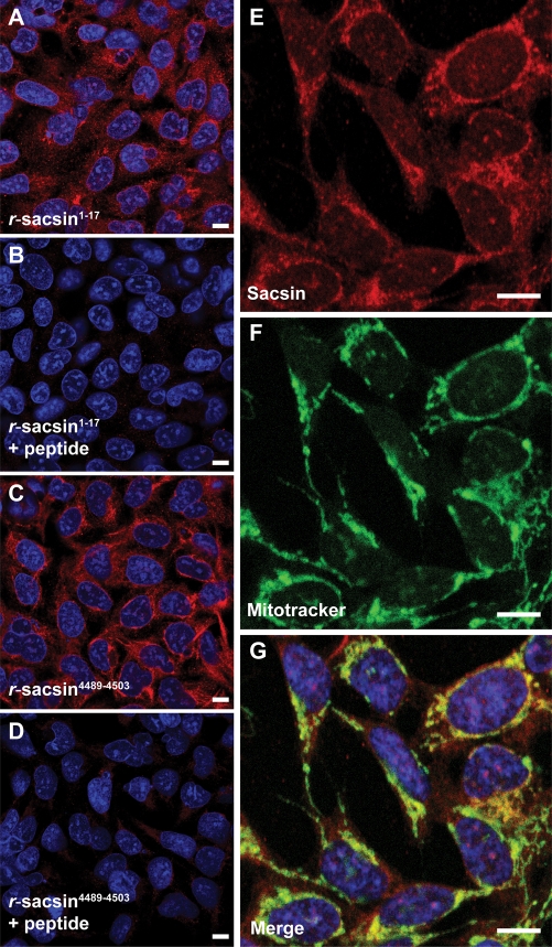
Figure 2.
Sacsin has a predominantly cytoplasmic localization with partial mitochondrial overlap. Immunofluorescent detection of sacsin in SH-SY5Y cells using r-sacsin1–17 (A, B) and r-sacsin4489–4503 (C, D). The antibodies were competed with immunizing peptide in (B and D). (E–G) Sacsin distribution (red) partially overlapped with a fluorescent mitochondria marker (green). Cells were imaged by laser scanning confocal microscopy. Microscope settings were constant for acquisition in the peptide competition experiments. Scale bar = 10 µm.

