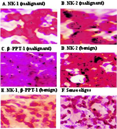
Figure 1.

In situ hybridization for PPT-I, NK-1, and NK-2 mRNA in breast biopsies. PPT-I, NK-1, and NK-2 mRNA was studied by in situ hybridization with tissues from paraffin-embedded breast biopsies. A representative stain from different benign (n = 21) and malignant (n = 25) tissues is shown at ×1,000 magnification. Tissues were hybridized with oligonucleotides specific for NK-1 (A), NK-2 (B and D), or β-PPT-I (C). E represents benign sections hybridized for NK-1 or β-PPT-I, and F shows the staining pattern with sense oligonucleotides from benign or malignant tissues.
