
Figure 1.
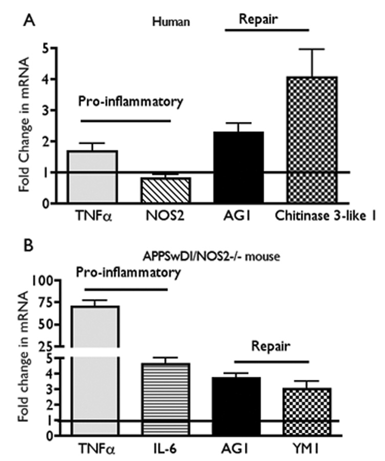
Panel A: Average fold change (± SEM) in mRNA levels of pro-inflammatory (TNFα; NOS2) and repair [arginase 1 (AG1) and chitinase 3-like-1 (CH3L1)] cytokines from mixed cortical lysates from humans with AD (average age approximately 78 yrs; Stage 4–5 Braak scale). Quantitative RT-PCR data from AD samples were compared to brain samples from aged matched normal individuals. De-identified autopsied brain samples were provided by the Kathleen Bryan Brain Bank at Duke University Medical Center, Durham, NC as described [30].
Panel B: Average fold change (± SEM) in mRNA from aged (52–54 week old) APPSwDI/NOS2−/− mice compared to aged matched wild type (WT) mice. Pro-inflammatory genes were TNFα and IL-6 and repair genes were AG1 and YM1 (mouse homolog for CH3L1). All values are significantly greater than control with p ≤ 0.05 (Student’s t test) (for humans, n = 29 control; AD; mixed gender; for mice n = 5–6 mice, mixed gender).