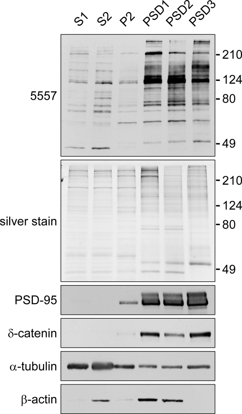
Fig. 2.
Enrichment of putative MAPK-phosphorylated proteins in postsynaptic density fractions. Shown is an immunoblot of rat brain fractions with MAPK phosphomotif antibody 5557. Brain extracts were fractionated by discontinuous sucrose gradient and detergent extraction into postnuclear supernatant (S1), cytosol plus light membranes (S2), crude synaptosomal fraction (P2), and sucrose gradient-purified synaptosomes extracted once or twice with 0.5% Triton X-100 (PSD1 and PSD2, respectively) or extracted once with 0.5% Triton X-100 followed by 3% Sarkosyl extraction (PSD3). Fractions (5 μg of protein) were immunoblotted with the indicated antibodies or visualized by silver stain. Phosphatase inhibitors were present throughout the PSD fractionation, hence the slightly different pattern compared with Fig. 1C.

