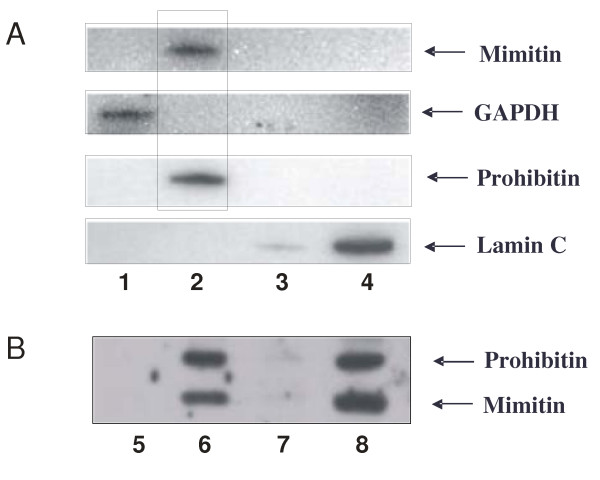
Figure 4.
Subcellular localization of mimitin. Subcellular fractionation of HepG2 cells was done with Qproteome Kit (A) and the obtained cytosolic (lane 1), membrane (lane 2), nuclear (lane 3) and cytoskeletal proteins (lane 4) were checked for the presence of appropriate markers with monospecific antibodies. Western blotting showed the presence of mimitin exclusively in the membrane fraction. In additional experiments cytosolic (lane 5), mitochondrial (lane 6), microsomal (lane 7) and crude nuclear fraction containing unlysed cells (lane 8), were isolated (see Materials and Methods). This result confirmed the presence of mimitin solely in mitochondria.

