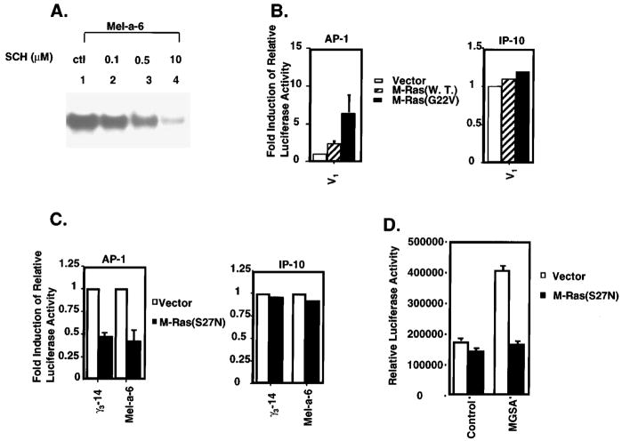
Figure 5.
Ras mediated AP-1 activation. (a) Ras inhibitor (SCH44342) blocks AP-1 binding activity: Mel-a-6 cells were pre-treated with the indicated concentrations of SCH44342 (lanes 2 – 4) or the solvent control (lane 1) overnight. Nuclear proteins were extracted and EMSA’s were performed by using 5 μg of protein and 0.4 ng of 32P-labeled AP-1 consensus oligonucleotide in EMSA. (b) M-Ras increases basal activation of AP-1: Left panel: V1 cells were cotransfected with 2.5 μg AP-1 luciferase reporter and either 5 μg pBK-CMV vector (empty bar), M-Ras (striped bar) or the dominant active form of M-Ras (G22V) (solid bar), together with 2.5 μg pBL-CAT3 plasmid. Luciferase and CAT activity were measured 48 h later. The relative luciferase activity represents the luciferase activity normalized by CAT activity. The results are reported as a mean (±s.e.m.) of fold-induction (the relative luciferase activity of dominant active form of M-Ras divided by the relative luciferase activity of pBK-CMV vector) from three independent experiments. Right panel: V1 cells were cotransfected with 2.5 μg IP-10 CAT reporter and 5 μg pBK-CMV vector (empty bar), M-Ras (striped bar) or the dominant active form of M-Ras (G22V) (solid bar), together with 2.5 μg pSV-β-Gal expression plasmid. CAT and Luciferase activity were measured 48 h later. (c) Dominant negative M-Ras blocks MGSA/GRO-increased basal AP-1 activation: Left panel: γ3-14 or Mel-a-6 cells were cotransfected with 2.5 μg AP-1 luciferase reporter and 5 μg pBK-CMV vector (empty bar) or the dominant negative M-Ras (solid bar), together with 2.5 μg pBL-CAT3 plasmid. Luciferase and CAT activity were measured 48 h later. The relative luciferase activity represents the luciferase activity normalized by CAT activity. The results are reported as a mean (±s.e.m.) of relative inhibition (the relative luciferase activity of dominant negative form of M-Ras divided by the relative luciferase activity of pBK-CMV vector) from three independent experiments. Right panel: γ3-14 or Mel-a-6 cells were cotransfected with 2.5 μg IP-10 CAT reporter and 5 μg pBK-CMV vector (empty bar) or the dominant negative M-Ras (solid bar), together with 2.5 μg pSV-β-Gal expression plasmid. CAT and Luciferase activity were measured 48 h later. (d) Dominant negative M-Ras blocks MGSA/GRO-induced AP-1 activation in V1 cells: V1 cells were cotransfected with 2.5 μg AP-1 luciferase reporter and 5 μg pBK-CMV vector (empty bar) or the dominant negative M-Ras (solid bar), together with 2.5 μg pBL-CAT3 plasmid. After transfection, cells were treated with carrier buffer alone or with 50 ng/ml MGSA/GROα for 48 h. The relative luciferase activity represents the luciferase activity normalized by CAT activity. The results are reported as a mean (±s.e.m.) of relative luciferase activity from three independent experiments

