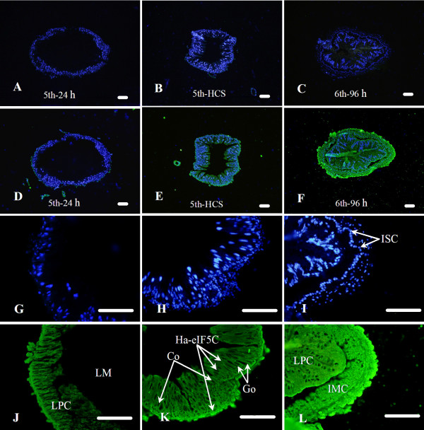
Figure 8.
Immunocytochemical localization of eIF5C in the midgut. Panels A-C are negative controls with pre-immune rabbit serum; panels D-F are midgut from feeding 5th instar larva (5th-24 h), molting 5th instar larva (5th-HCS) and 6th-96 h (wandering) larva; panels G and J, H and K, I and L are the magnified D, E, F, respectively; nuclear staining was done by DAPI (G, H, I) and the positive signals were detected by ALEXA 488 assay (J, K, L), panels A-F are overlay. LM, lumen of midgut; LPC, larval polyploidy cells; ISC, intestinal stem cell; IMC, imaginal cells; Co, clumnar cells; Go, goblet cells. Scale bar = 100 μm.

