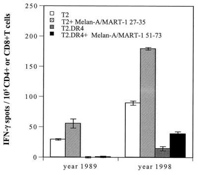
Figure 1.
Recognition of the Melan-A/MART-127–35 and Melan-A/MART-151–73 peptides by CD8+ or CD4+ T cells, respectively, derived from peripheral blood of patient UPCI-MEL 136. CD8+ and CD4+ T cells were isolated from cryopreserved PBMC of patient UPCI-MEL 136 collected in 1989 and 1998, seeded at 105 per well, and tested for reactivity against, respectively, T2 cells pulsed with the Melan-A/MART-127–35 peptide or T2.DR4 cells pulsed with the Melan-A/MART-151–73 peptide. After a culture period of 20 h at 37°C, IFN-γ spots were developed and counted by computer-assisted video image analysis. Each bar represents the mean spot number of triplicates ± SD with 105 CD8+ T cells (screened against T2 or T2 + Melan-A/MART-127–35 peptide) or CD4+ T cells (screened against T2.DR4 or T2.DR4 + Melan-A/MART-151–73 peptide) initially seeded per well. T2 cells pulsed with HLA-A2-restricted peptides (including tyrosinase1–9 and tyrosinase369–377D) or T2.DR4 cells pulsed with HLA-DR4-restricted peptides (including tyrosinase56–70, gp10044–59, gp100167–189, Melan-A/MART-143–57, or Melan-A/MART-1102–116) also were tested in this assay: no immunoreactivity against these peptides could be detected (data not shown).

