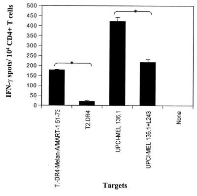
Figure 3.

Recognition of the Melan-A/MART-1 51–73 peptide and UPCI-MEL 136.1 cell line by CD4+T cells in patient UPCI-MEL 136. CD4+ T cells were isolated from the peripheral blood of patient UPCI-MEL 136 and stimulated in vitro with autologous DC pulsed with the Melan-A/MART-1 peptide as described in Materials and Methods. Ten thousand CD4+ T, obtained after five rounds of in vitro stimulation, were incubated in a 20-h IFN-γ ELISPOT assay in the presence of T2.DR4 cells pulsed with the Melan-A/MART-151–73 peptide (1 μg/ml), UPCI-MEL 136.1 cells +/− anti-HLA-DR antibodies (L243), or UPCI-MEL 136.1 cells +/− anti-HLA-A,B,C antibodies (W6/32, data not shown). Data from one representative experiment of six performed is depicted. * indicate significant results, i.e., P < 0.05. The mean values of experimental and background counts were compared by using Student's unpaired t test.
