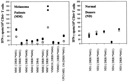
Figure 6.
Basal CD4+ T cell recognition of the Melan-A/MART-151–73 peptide in 10 HLA-DR4+ patients with melanoma. CD4+ T cells were isolated from cryopreserved PBMC of 10 DR4+ melanoma patients (A) or five DR4+ normal donors (B), seeded at 105 per well in duplicate, and tested for reactivity against T2.DR4 cells pulsed with the Melan-A/MART-151–73 peptide or unpulsed T2.DR4 cells, respectively. After a culture period of 20 h at 37°C, IFN-γ spots were developed and counted by computer-assisted video image analysis. Each symbol represents the spot number observed in one individual well of the ELISPOT assay with 105 CD4+ T cells initially seeded per well. Each circle represents the spot number obtained in the presence of unpulsed T2.DR4 cells (○) or Melan-A/MART-151–73-pulsed T2.DR4 cells (●), respectively. Of note, the T cell responses directed against unpulsed T2.DR4 cells displayed significant donor variations, consistent with responder T cell recognition of Epstein–Barr virus-derived epitopes expressed by T2.DR4 cells, as reported (17). The data were obtained from one representative experiment of two performed, with comparable results observed in both assays.

