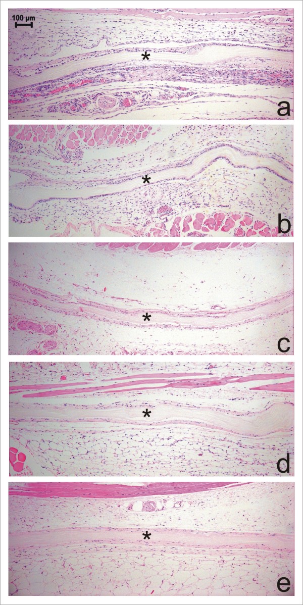
Figure 3.
Histomorphology of a microbial cellulose membrane implanted subcutaneously in mice and surrounding tissue reaction 7(a), 15(b), 30(c), 60(d) and 90(e) days postoperatively. Observe the presence of the intact membrane (*) surrounded by immature granulation tissue and newly formed vessels and capillaries (a). At 15 days post-surgery, a reduction in inflammatory infiltrate, especially of lymphocytes, is observed (b). At 30 days postoperatively observe the collagen fibers commencing orientation parallel to the implant's surface (c). No inflammatory infiltrate is observed and the connective tissue surrounding the membrane is mature at 60 (d) and 90 (e) days post-surgery. (HE, Obj. ×10).

