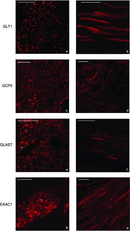
Fig. 3.
Immunofluorescence analysis of the sciatic nerve with affinity-purified primary antibody against GLT1, GCPII, GLAST and EAAC1. In transverse and longitudinal sections of the sciatic nerve (a,b), GLT1 immunolabelling was stronger in the outer cytoplasm of Schwann cells around nerve fibres (arrows). In some cases, the axon also showed positive, although weak, immunoreactivity for anti-GLT1 antibodies (a). In transverse section of the sciatic nerve (c), GCPII immunolabelling was confined to the area around axons, probably in the outer cytoplasm of Schwann cells (arrow). In longitudinal section (d), anti-GCPII immunoreactivity was localized only on the surface of the nerve fibres with no axonal labelling. Anti-GLAST and anti-EAAC1 immunoreactivities (e,f and g,h) were present in the myelin layer immediately around the axon. As shown by the arrows in (e) and (g), spots of strong labelling were visible on the external periphery of the myelin itself. Bar: 50 µm.

