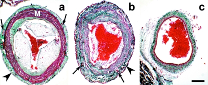
Fig. 3.
Transverse sections (see also Fig. 2) of the conus arteriosus (a), distal component of the cardiac outflow tract (b) and ventral aorta (c) of an adult Galeus atlanticus. The thickness of the distal component wall is similar to that of the conus arteriosus; the aorta has a thinner wall. The walls of both the conus and the distal component of the outflow tract are covered by epicardium (arrowheads) and crossed by coronary arteries (arrows). M, myocardium. Collagen is stained green. Masson-Goldner's trichrome. Scale bar = 300 μm.

