Figs 4 and 5.
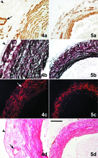
Tranverse sections of the distal component (DC) of the cardiac outflow tract (4) and ventral aorta (5). (4a) The DC wall has a thick layer of smooth muscle cells (brown labelling) forming concentric sheets. (5a) The aortic media shows several densely arranged smooth muscle layers. (4b) In the DC, elastin (black staining) forms equidistant concentric sheets that alternate with the smooth musculature. Elastin is very scarce in the subepicardial space. (5b) Elastin is abundant in the aortic media and scattered in the adventitia. (4c) Staining plus polarization microscopy enhance the natural birefringence of the collagen bundles, which follow a more or less concentric pattern through the whole wall of the DC wall and are more abundant in the subepicardial space. (5c) Collagen bundles are orientated longitudinally in the aortic media; in the adventitia they are more abundant forming a meshwork. (4d) Glycoconjugates (magenta) follow the pattern of distribution of elastin in the DC wall. (5d) Glycoconjugates follow the pattern of distribution of elastin in the aorta. (a) α-actin immunolabelling; (b) Orcein-HCl staining; (c) Picrosirius staining plus polarization microscopy; (d) PAS. The arrowhead marks the epicardium. The arrow points to a coronary artery located in the subepicardial space. Scale bars = a, b, d, 100 μm; c, 200 μm.

