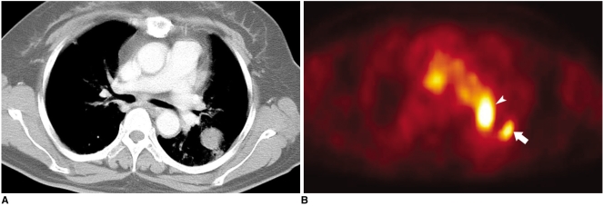
Fig. 3.
Tuberculoma in a 53-year-old female.
A. Contrast-enhanced CT scan shows a round mass in the left lower lobe.
B. Axial FDG-PET image shows intense uptake (arrow) in the left upper lobe suggesting a malignant condition with a maximum standardized uptake value of 4.3. The pathologic examination reveals tuberculoma. Another lesion showing high FDG uptake (arrowhead) is a pulmonary artery.

