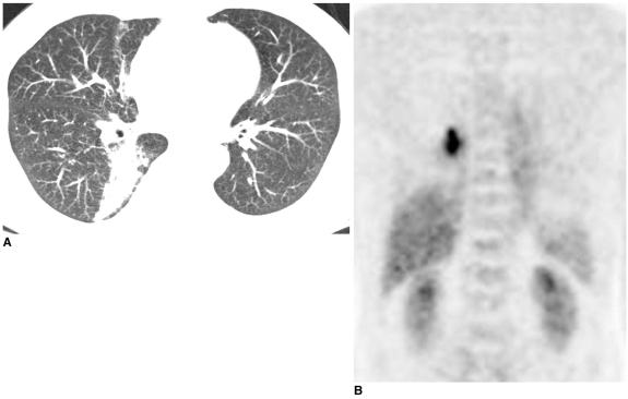
Fig. 7.
Paragonimiasis in a 68-year-old male.
A. In the lung window setting, axial transverse CT scan shows linear, wedge shaped consolidation and small centrilobular nodules. Radiologic diagnosis is atypical tuberculosis.
B. Coronal section of FDG-PET image shows intense uptake in the right lower lung zone. Bronchoscopic washing and sputum cytology reveals many parasitic eggs of paragonimus species.

