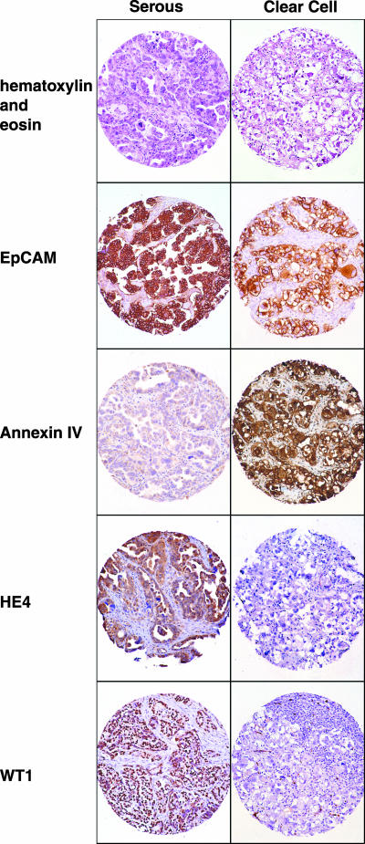
Figure 5.
Immunohistochemistry: ovarian cancer tissue arrays comprised of both serous (left panel) and clear cell (right panel) ovarian cancers. Hematoxylin and eosin staining is shown in the top panel. Staining of a representative case of serous and clear cell, respectively, were stained with antibodies against EPCAM, annexin IV, HE4, and WT1.

