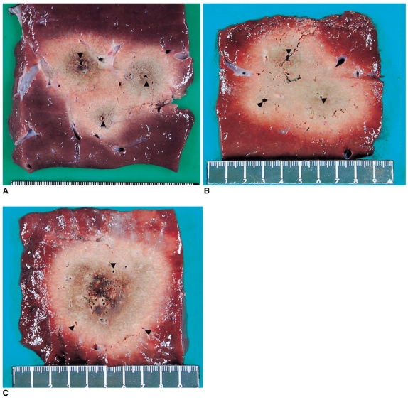
Fig. 3.
Comparison of radiofrequency-induced coagulation created by applying radiofrequency in the consecutive, simultaneous and multipolar modes with a 4-cm inter-probe distance. Note that the mean short-axis diameter was largest in the bipolar mode. The arrowheads indicate the electrode insertion sites.
A. Cut section of the specimen created with the consecutive monopolar RFA shows three separate ablation spheres.
B. Cut section of the specimen created with the simultaneous monopolar RFA. The long-axis and short axis diameters of the ablation zone were 5.5 cm and 3.8 cm, respectively.
C. Cut section of the specimen created with the bipolar RFA. The long-axis and short axis diameters of the ablation zone were 5.5 cm and 5.1 cm, respectively.

