Fig. 5.
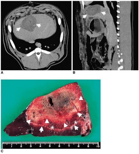
Contrast-enhanced CT scans and photographs of the liver treated with multipolar RFA for 20 minutes in an in vivo pig model.
A. Axial contrast-enhanced CT scan obtained 4 days after multipolar RFA reveals a focal non-enhanced region (white arrows) in the liver.
B. Sagittal reformatted image shows a well-defined non-enhanced region (white arrows) in the liver.
C. Gross hepatic section staining with 2% 2,3,5,-triphenyl tetrazolium chloride showing a central white ablation zone without staining (arrowheads), and a peripheral partial staining zone (*) surrounded by a white rim (arrows). The microscopic slide shows a confined area of coagulation necrosis surrounded by a peripheral hemorrhagic zone consisting of necrotic hepatocytes, interstitial hemorrhage and leukocyte infiltrates (not shown).

